medial calcaneal tubercle, check these out | What is the medial calcaneal tubercle?
By Sarah Rowe
What is the medial calcaneal tubercle?
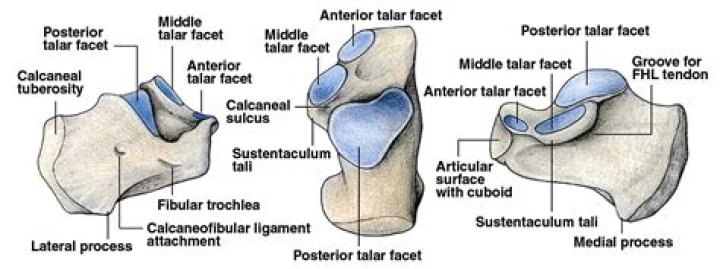
On the lateral side is commonly a tubercle called the calcaneal tubercle (or trochlear process). This is a raised projection located between the tendons of the peroneus longus and brevis. It separates the two oblique grooves of the lateral surface of the calcaneus (for the tendons of the peroneal muscles).
Where is the medial calcaneal tubercle?
The plantar surface of the calcaneal tuberosity projects forward on the plantar surface as a medial (larger) and lateral (smaller) process and at its most anterior projection is the calcaneal tubercle, where the short plantar ligament attaches.
What attaches to the medial calcaneal tubercle?
There is a prominent medial margin on the medial process of the calcaneal tuberosity which provides attachments for the superficial part of the flexor retinaculum and distally the plantar aponeurosis. Some muscles also attach there including abductor hallucis and flexor digitorum brevis.
What is the purpose of calcaneal tuberosity?
The inferior surface (plantar surface) of calcaneus is bounded posteriorly by a transverse elevation, the calcaneal tuberosity,which is depressed in the middle and prolonged at either end into a process; the lateral process, small, prominent, and rounded, gives origin to part of the Abductor digiti quinti; the medial
Where is medial heel pain?
Usually, the heel pain overrides pain through the arch of the foot, more distally. If you are suffering with medial heel pain, then you will probably notice that the pain is very apparent first thing in the morning when your foot hits the floor. This is very common and very typical of plantar fasciitis.
What is the inner heel called?
In humans the heel consists of the calcaneus (largest of the tarsal bones), cushioned below by a bursal sac, fat pad, and thickened skin. The calcaneus is roughly rectangular, articulating above with the talus bone of the ankle joint and in front with the cuboid, another tarsal bone.
How do you treat Baxter’s nerve entrapment?
Podiatry treatment for Baxter’s nerve entrapment
Foot strapping.Orthotics to address foot mechanics.Stretching and strengthening programs.Footwear advice and modification.Rest, ice and activity modification.Oral medications (such as NSAIDs)Ultrasound therapy and heat.
Does Achilles tendonitis go away?
With rest, Achilles tendonitis usually gets better within 6 weeks to a few months. To lower your risk of Achilles tendonitis again: Stay in good shape year-round.
How do you fix nerve pain in your heel?
Home-based treatments for the condition include applying ice, taking anti-inflammatory medications, and stretching the foot daily. Your doctor may be able to ease pain with corticosteroid injections, physical therapy, orthotics, or surgery. Learn more about plantar fasciitis.
What tendon attaches to the calcaneus?
The Achilles tendon attaches to the calcaneal tubercle.
What structures attaches to the calcaneal tuberosity?
Attachments
Triceps surae, i.e. gastrocnemius and soleus (insertion: middle facet of posterior surface of calcaneus through calcaneal/Achilles tendon.Abductor hallucis (origin: the medial process of calcaneal tuberosity)Flexor digitorum brevis (origin: the medial process of calcaneal tuberosity and plantar aponeurosis)
What attaches to the sustentaculum tali?
Several ligamentous structures attach to the sustentaculum tali: plantar calcaneonavicular ligament (anterior surface) deltoid ligament (medial surface) medial talocalcaneal ligament.
Why is the sustentaculum tali important?
At the upper and forepart of the medial surface of the calcaneus is a horizontal eminence, the sustentaculum tali, which gives attachment to a slip of the tendon of the Tibialis posterior.
What is the navicular tuberosity?
The navicular tuberosity is an osseous prominence that arises on the medial aspect of the navicular bone. It is responsible for the insertion of plantar and medial navicular ligaments, and the posterior tibial tendon as well.
Why does my heel bone hurt?
Common causes of heel pain include obesity, ill-fitting shoes, running and jumping on hard surfaces, abnormal walking style, injuries and certain diseases. Plantar fasciitis is inflammation of the ligament that runs the length of the foot, commonly caused by overstretching.
How do you treat calcaneus pain?
How can heel pain be treated?
Rest as much as possible.Apply ice to the heel for 10 to 15 minutes twice a day.Take over-the-counter pain medications.Wear shoes that fit properly.Wear a night splint, a special device that stretches the foot while you sleep.Use heel lifts or shoe inserts to reduce pain.
What is the best exercise for heel pain?
Here are six exercises from physical therapists that you can try at home.
Plantar Fascia Massage. Note: You should not experience pain during this exercise. Heel Raise. Floor Sitting Ankle Inversion With Resistance. Seated Toe Towel Scrunches. Seated Plantar Fascia Stretch. Wall-Facing Calf Stretch.
What causes medial plantar nerve entrapment?
When there is repetitive impact to the abductor halluces muscle, such as during long distance running, the muscle can become swollen and inflamed. This then presses against the medial plantar nerve and causes the nerve to be compressed or entrapped. This is what causes the pain.
Related Archive
More in general 
harry potter wizarding world japan, latest free online harry potter movies, best HD videos you should watch in 2022 – 2023

harry potter vs voldemort in the deathly hallows, latest free online harry potter movies, best HD videos you should watch in 2022 – 2023