lateral pterygoid action, check these out | What is the function of the lateral pterygoid plate?
By Mia Kelly
Function. The primary function of the lateral pterygoid muscle is to pull the head of the condyle out of the mandibular fossa along the articular eminence to protrude the mandible. A concerted effort of the lateral pterygoid muscles helps in lowering the mandible and opening the jaw.
What is the function of the lateral pterygoid plate?
The lateral pterygoid allows the jaw to move in a horizontal direction during mastication (chewing).
What do the pterygoid muscles do?
The pterygoid muscles are two of the four muscles of mastication, located in the infratemporal fossa of the skull. The primary function of the pterygoid muscles is to produce movements of the mandible at the temporomandibular joint.
What is the action of the medial pterygoid?
The medial pterygoid muscle attaches to the angle of the mandible and to the lateral pterygoid plate to form a sling with the masseter muscle that suspends the mandible (Figure 6-19). The primary action is to elevate the mandible and laterally deviate it to the opposite side.
What movements are performed when the lateral pterygoid muscle is contracted?
The unilateral contraction of the lateral pterygoid muscle with the ipsilateral medial pterygoid muscle results in lateral mandibular movement to the contralateral side. This movement is observable during functional and parafunctional lateral excursive movements, i.e., during chewing stroke, masticating, and clenching.
What are structures of pterygoid process?
Each pterygoid process projects inferiorly from the junction of the body and greater wing of the sphenoid bone and bifurcates into a medial pterygoid plate and a lateral pterygoid plate. At the inferior tip of the medial pterygoid plate is the small hook-shaped process, the pterygoid hamulus.
Which are functions of the medial and lateral pterygoid muscles quizlet?
Two-headed, fan-shaped muscle located in the infratemporal fossa of the skull. It is one of the four masticatory muscles, along with the medial pterygoid, temporalis and masseter muscles. All these muscles act upon the temporomandibular joint (TMJ) to enable chewing (mastication) and biting.
Why is the lateral pterygoid called the peripheral heart?
The pterygoid muscles and other muscles of mastication pump the blood from this plexus and are considered a “peripheral heart”.
What is the function of Digastric muscle?
Structure and Function
The digastric muscle functions during swallowing, chewing, and speech. The anterior belly of the digastric is one of the three suprahyoid muscles which stabilizes the hyoid during swallowing, an action critical in protecting the airway while eating.
Can you palpate the lateral pterygoid?
The lateral pterygoid muscle is inevitably quite tender in most individuals with TMJ issues or bruxism. To palpate, place the index finger inside the mouth. Apply pressure in a cranial direction just underneath the zygomatic arch.
What is protrusion movement?
Protrusion involves a movement going straight ahead or forward. Retrusion is the opposite and involves going backwards. Anatomical structures capable of such actions are the tongue, chin (mandible) and lips.
How do you examine lateral pterygoid?
Attempted palpation of what has been thought to be this structure is commonly done by placing the forefinger, or the little finger, over the buccal area of the maxillary third molar region and exerting pressure in a posterior, superior, and medial direction behind the maxillary tuberosity (Figure 2).
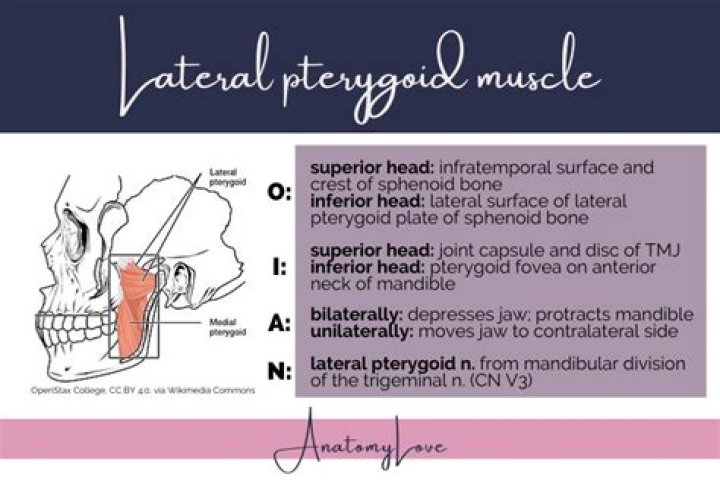
Where is the lateral pterygoid muscle?
Lateral pterygoid is a two-headed, fan-shaped muscle located in the infratemporal fossa of the skull. It is one of the four masticatory muscles, along with the medial pterygoid, temporalis and masseter muscles.
Where are the Pterygoid process found?
Two pairs of bony plates, the pterygoid processes, arise from the base of each alisphenoid bone. The outer plates are nearly horizontal in position. They extend from the posterior end of the maxillary bone caudad and laterad to the lateral surface of the tympanic bulla.
Related Archive
More in updates 
harry potter wizards unite mod joystick, latest free online harry potter movies, best HD videos you should watch in 2022 – 2023

harry potter vs voldemort poster, latest free online harry potter movies, best HD videos you should watch in 2022 – 2023