iac brain, check these out | What is the IAC in the brain?
By Liam Parker
What is the IAC in the brain?
Magnetic resonance imaging (MRI) of the internal auditory canal (IAC) is a non-invasive, painless diagnostic imaging procedure that uses using radio waves and a strong magnetic field to create detailed images of the bony canal that transmits nerves and blood vessels from the base of the brain to the inner ear.
What is IAC in radiology?
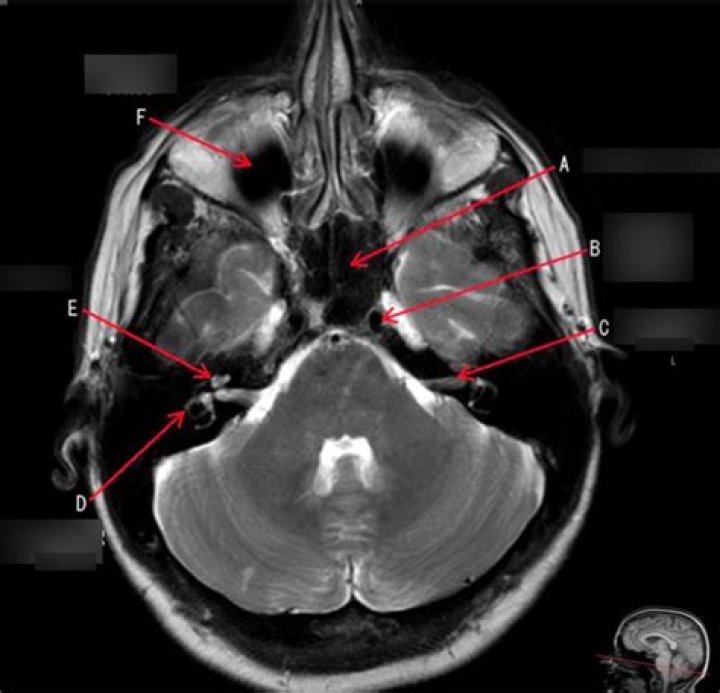
The internal acoustic canal (IAC), also known as the internal auditory canal or meatus (IAM), is a bony canal within the petrous portion of the temporal bone that transmits nerves and vessels from within the posterior cranial fossa to the auditory and vestibular apparatus.
What can an IAC MRI show?
Common applications of MRI include diagnostic evaluation of sensorineural hearing loss, assessment of cochlear implant candidacy, monitoring for residual or recurrent cholesteatoma within the tympanomastoid space, and monitoring for vestibular schwannoma within the inner auditory canal or cerebellopontine angle.
What is an IAC lesion?
IAC meningoceles are rare lesions which have not been well described in the literature. The presence of an IAC meningocele can result in compression of the 7–8th nerve, and the lack of a bone covering between the IAC and cochlea can have implications for hearing and facial-nerve function.
Why do I need an MRI scan for hearing loss?
An MRI scan may reveal a growth on the nerve pathway that connects the ear to the brain, such as an acoustic neuroma. These growths can prevent the ear from functioning well and may cause hearing loss.
What can an MRA detect in the brain?
An MRA of the head is done to look at the blood vessels leading to the brain to check for a bulge (aneurysm), a clot, or a narrowing (stenosis) because of plaque.
Does MRI brain include IAC?
Magnetic resonance imaging (MRI) is presently the study of choice for assessment of the internal auditory canal (IAC). MRI provides excellent assessment of the IAC and the bony changes occurring in the canal walls, and it provides excellent demonstration of the content of the canal.
Can an MRI scan detect inner ear problems?
MRI scans use a magnetic field and radio waves to create computerized, three-dimensional images of the ear and the nerve that carries signals from the inner ear to the brain. An MRI scan may reveal a buildup of fluid or inflammation in the inner ear or a growth on the nerve.
Is MRI IAC with contrast?
Objective: Non-contrast MRI of the internal auditory canal (IAC) using high-resolution T2WI (T2 weighted image) has been proposed as the primary screening study in patients with sudden or asymmetric sensorineural hearing loss (ASNHL).
What should you not do before an MRI?
The absolute most important thing not to do before an MRI is to lie or leave out information when talking to your doctor or the MRI technicians. MRIs are not safe for certain people. If you are pregnant or breastfeeding, you may be asked to delay the MRI, if possible.
What is Cerebellopontine?
The cerebellopontine angle (CPA) (Latin: angulus cerebellopontinus) is located between the cerebellum and the pons. The cerebellopontine angle is the site of the cerebellopontine angle cistern one of the subarachnoid cisterns that contains cerebrospinal fluid, arachnoid tissue, cranial nerves, and associated vessels.
What nerves are affected by an acoustic neuroma?
Acoustic neuromas are noncancerous, usually slow growing tumors that form along the branches of the eighth cranial nerve (also called the vestibulocochlear nerve). This nerve leads from the brain to the inner ear and branches into divisions that play important roles in both hearing and balance.
Are schwannomas painful?
Symptoms of a schwannoma may be vague and will vary depending on its location and size, but may include a lump or bump that can be seen or felt, pain, muscle weakness, tingling, numbness, hearing problems, and/or facial paralysis. Sometimes schwannomas do not cause any symptoms.
Is hearing loss a neurological disorder?
Many studies have revealed that neurological disorders manifest with hearing loss, in addition to typical nervous symptoms. The prevalence, manifestations, and neuropathological mechanisms underlying vary among different diseases.
What are the symptoms of a tumor in the ear?
Signs of an ear tumor include:
Dizziness or balance problems.Ear bleeding or discharge.Ear pain.Headaches.Hearing loss.Nonhealing wound or sore.Skin discoloration, new moles or changes to a mole.Swollen lymph nodes.
Is tinnitus a symptom of brain tumor?
Symptoms that may indicate a possible cranial base tumor include: Headaches or dizziness. Tinnitus (ringing in the ear) Difficulty breathing.
Related Archive
More in updates 
harry potter wizards unite mod joystick, latest free online harry potter movies, best HD videos you should watch in 2022 – 2023

harry potter vs voldemort poster, latest free online harry potter movies, best HD videos you should watch in 2022 – 2023