
dipnech life expectancy, check these out | Is DIPNECH a cancer?
By James Austin
Is DIPNECH a cancer?
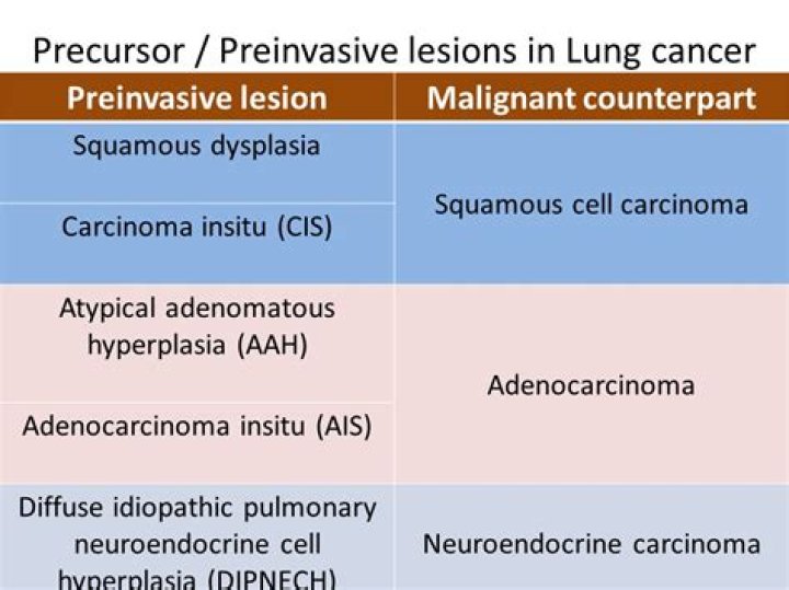
DIPNECH is recognised by the 2015 World Health Organization (WHO) classification of lung tumours as a pre-neoplastic lesion [5]. In fact, while overall there are insufficient well-documented data to support it as a pre-neoplastic condition, DIPNECH is generally thought of as a precursor for malignancy.
How is DIPNECH treated?
Currently, the management and treatment options for DIPNECH have included clinical observation, oral and inhaled steroids, chemotherapy, surgical lung resection, and even lung transplantation [3,4,9,10].
How common is DIPNECH?
DIPNECH remains a rare disease more commonly diagnosed in women in their early 60’s. DIPNECH appears to have an indolent course with obstructive symptoms being the most common finding. A minority of patients experienced symptom relief with therapy.
How many cases of DIPNECH are there?
Diffuse idiopathic pulmonary neuroendocrine cell hyperplasia (DIPNECH) is a rare clinical condition with only about 100 cases reported in the literature.
Is DIPNECH fatal?
The clinical course however, is characterized by slowly progressive, unspecific pulmonary symptoms such as long-lasting dry cough, wheezing and exertional dyspnoea [2,4-8]. Nonetheless, DIPNECH can also cause severe airflow obstruction and respiratory failure which can prove fatal [2,3,7,9].
What causes DIPNECH?
The cause of DIPNECH is still unknown. During fetal development, pulmonary neuroendocrine cells (PNC) are located throughout the whole respiratory tract as they have a key role in the regulation of lung development. In adulthood, PNCs are typically less numerous.
Is DIPNECH genetic?
Only 40 cases of DIPNECH have been reported in the literature to date [1] and there are no predictive histological or genetic data available so far. However, it has become generally accepted that DIPNECH is a precursor to pulmonary carcinoid tumors [3].
Is a typical carcinoid tumor malignant?
Typical carcinoids and atypical carcinoids are, respectively, low- and intermediate-grade neuroendocrine tumors. Approximately 80% of pulmonary carcinoids occur centrally, and 20% are peripheral. All bronchial carcinoids are malignant and have the potential to metastasize.
What is Nehi lung disease?
Neuroendocrine cell hyperplasia of infancy (NEHI), initially described as persistent tachypnea of infancy, is a rare lung disease first defined in 2005 [1]. The etiology is unknown, but genetic mechanisms may play a role.
What is hyperplasia lung?
Pulmonary hyperplasia is a serious pathological event which occurs in neonatal medicine. It leads to pulmonary hypertension and acute respiratory distress syndrome. Histologically the lungs classically exhibit “synchronous” hypermaturity”.
What is atypical adenomatous hyperplasia?
Atypical adenomatous hyperplasia of the lung (AAH) is defined as a peripheral focal proliferation of atypical cuboidal or columnar epitheial cells along the alveoli and respiratory bronchioles (1).
What are carcinoid Tumorlets?
Carcinoid tumorlets are defined as hyperplasia of neuroendocrine cells that are 5 mm or less in size and lack of mitotic activity and necrosis.
What foods should be avoided with carcinoid syndrome?
The following foods and/or eating habits are often triggers and may make these symptoms worse:
Large meals.High fat meals.Alcohol.Spicy foods.Raw tomatoes.Foods containing moderate or high amounts of amines (please see the list on page # 3)
Can you see carcinoid tumors on a CT scan?
Chest X-ray, computed tomography (CT) scan, and magnetic resonance imaging (MRI) scan are all useful in diagnosis. OctreoScan. This is a special type of scan that is most often used to find carcinoid tumors. This scan is taken after injection of a radioactive substance that is picked up by carcinoid tumor cells.
Related Archive
More in news 
harry potter x reader first time, latest free online harry potter movies, best HD videos you should watch in 2022 – 2023

harry potter trivia kansas city, latest free online harry potter movies, best HD videos you should watch in 2022 – 2023