c arm full form, check these out | What is C-arm used for?
By Andrew Walker
CARM Stands For : Computer Assisted Radio Monitoring.
What is C-arm used for?
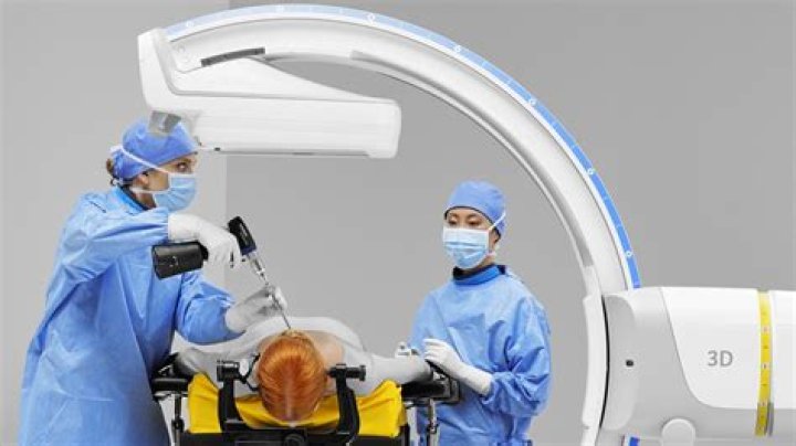
A mobile C-arm is a medical imaging device that is based on X-ray technology and can be used flexibly in various ORs within a clinic. The name is derived from the C-shaped arm used to connect the X-ray source and X-ray detector to one another.
What is C-arm in surgery?
C-Arm is a mobile imaging unit used primarily for fluoroscopic imaging during surgical and orthopedic procedures. It also consists of a computer workstation used to view, manipulate, store and transfer the images.
Who invented C-arm?
1955 Prior to 1955, X-ray systems were unable to change direction. Philips therefore developed the first C-arm – an X-ray system in the form of a half moon.
What is difference between C-arm and fluoroscopy?
The C-arm machine is a fluoroscopy system. Fluoroscopy is a method providing real-time X-ray imaging, which is particularly useful for guiding various diagnostic and interventional procedures. Though you should remember that C-arms are generally not used in diagnostics, they are made for surgery.
What is the difference between C-arm and O-arm?
O-arm navigation system use is shorter in radiation time and larger in radiation exposure than C-arm fluoroscopy navigation system. However, the amount of the radiation exposure per unit time in O-arm navigation system is larger than in C-arm fluoroscopy navigation system.
What is a mini C-arm?
Mini C-arm is a mobile fluoroscope with less radiation exposure to the surgeon, patient and theatre personnel. It is used for intra-operative imaging of a host of procedures and its simplicity of use, low cost and compact nature make it popular for routine use.
How much radiation do you get from C-arm?
With ionising radiation produced by a standard C-arm, this is roughly equal to equivalent doses of 0.113 μSv per lateral image and 0.043 μSv per PA image 16.
What is fluoroscopy imaging?
Fluoroscopy is a study of moving body structures–similar to an X-ray “movie.” A continuous X-ray beam is passed through the body part being examined. The beam is transmitted to a TV-like monitor so that the body part and its motion can be seen in detail.
How do you use fluoroscopy?
During a fluoroscopy procedure, an X-ray beam is passed through the body. The image is transmitted to a monitor so the movement of a body part or of an instrument or contrast agent (“X-ray dye”) through the body can be seen in detail.
What is the purpose of the image intensifier?
The function of the x-ray image intensifier in the fluoroscopic imaging system is to convert the x- ray spectrum transmitted through the patient into a highly visible image.
Where is the image intensifier on a C-arm?
Fixed systems may have a c-arm mounted to a ceiling gantry, with a separate control area. Most systems arranged as c-arms can have the image intensifier positioned above or below the patient (with the X-ray tube below or above respectively), although some static in room systems may have fixed orientations.
Related Archive
More in updates 
harry potter wizards unite mod joystick, latest free online harry potter movies, best HD videos you should watch in 2022 – 2023

harry potter vs voldemort poster, latest free online harry potter movies, best HD videos you should watch in 2022 – 2023